Anatomy & Physiology Lab Manual Answers: A Comprehensive Plan
This resource provides comprehensive lab solutions, supporting Anatomy & Physiology I at the University of Georgia, utilizing OER grants for enhanced student learning․
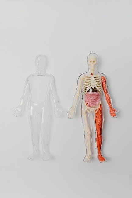
Lab 1: Organ Systems and Body Organization
This initial lab focuses on establishing a foundational understanding of the human body’s intricate organization․ Students will explore the eleven major organ systems – integumentary, skeletal, muscular, nervous, endocrine, cardiovascular, lymphatic, respiratory, digestive, urinary, and reproductive – and their interconnected functions․ The lab emphasizes levels of structural organization, progressing from chemical to organismal levels, providing a holistic view of bodily systems and their collaborative roles in maintaining life․
Overview of Human Organ Systems
A detailed exploration of eleven vital systems forms the core of this lab․ Students will investigate the integumentary, skeletal, muscular, nervous, endocrine, cardiovascular, lymphatic, respiratory, digestive, urinary, and reproductive systems․ Emphasis is placed on understanding each system’s unique components, their integrated functions, and how they contribute to overall homeostasis within the human body, fostering a comprehensive anatomical perspective․
Levels of Structural Organization
The human body exhibits a hierarchical arrangement, progressing from simple to complex structures․ This lab examines the six levels: chemical, cellular, tissue, organ, organ system, and organismal․ Students will analyze how each level builds upon the previous one, demonstrating emergent properties and illustrating the interconnectedness essential for maintaining life and proper physiological function․
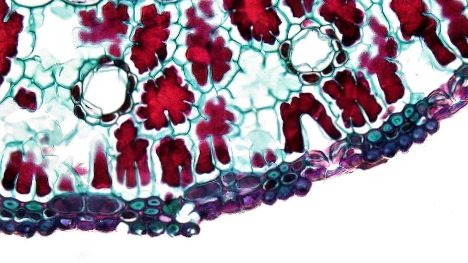
Lab 2: Microscopy – Exploring the Microscopic World
This lab introduces fundamental microscopy techniques crucial for visualizing anatomical structures beyond the naked eye’s capabilities․ Students will learn proper microscope component usage, slide preparation methods, and observation skills․ Mastering these skills is vital for understanding histology and cellular structures, forming the foundation for advanced anatomical and physiological investigations․
Microscope Components and Function
Understanding each component is key to effective microscopy․ This includes the ocular lens, objective lenses (varying magnification), stage, condenser, diaphragm, and illumination source․ Students will learn how each part contributes to image clarity, magnification, and resolution․ Proper adjustment of these components ensures optimal visualization of microscopic anatomical details for accurate analysis․
Preparing and Observing Slides
Effective slide preparation is crucial for clear microscopic observation․ This involves techniques like wet mounts, staining (to enhance contrast), and sectioning (for thicker specimens)․ Students will practice proper slide handling, focusing techniques, and identifying artifacts․ Careful observation and documentation of microscopic structures are essential skills developed through this lab component․
Lab 3: Tissues – The Building Blocks of Life
This lab focuses on the foundational tissues composing the human body․ Students will examine epithelial and connective tissues, identifying their distinct structures and correlating them with specific functions․ Histology, the study of tissues, is a core component, utilizing microscopy to differentiate tissue types․ Understanding tissue organization is vital for comprehending organ system function;
Epithelial Tissue Types and Functions
This section details the diverse world of epithelial tissues, categorized by shape and layering․ Students will learn to distinguish squamous, cuboidal, and columnar epithelia, alongside simple, stratified, and pseudostratified arrangements․ Key functions explored include protection, absorption, secretion, and excretion, relating structure directly to physiological roles within the body․
Connective Tissue Types and Functions
Connective tissues provide support, connection, and protection throughout the body․ This lab focuses on identifying various types – including connective tissue proper (loose & dense), cartilage, bone, and blood․ Students will analyze their unique cellular structures and extracellular matrices, correlating these features with functions like binding, transport, and structural support․
Lab 4: Integumentary System – Skin and its Appendages
This lab explores the skin’s structure and function, examining the epidermis and dermis layers․ Students will identify epidermal strata, dermal components like hair follicles and sweat glands, and analyze their roles in protection, sensation, and thermoregulation․ Detailed illustrations and photographs enhance understanding of this vital organ system․
Epidermis Structure and Layers
This section details the epidermis, the outermost skin layer, focusing on its distinct strata: basale, spinosum, granulosum, lucidum, and corneum․ Students will learn how keratinization occurs within these layers, contributing to skin’s protective barrier․ Microscopic observation aids in identifying cell types and understanding the epidermis’ role in vitamin D synthesis and immunity․
Dermis Structure and Components
The dermis, a deeper skin layer, is explored, emphasizing its papillary and reticular regions․ Students will identify collagen and elastic fibers providing strength and elasticity․ This lab focuses on dermal structures like blood vessels, nerves, hair follicles, and glands – sebaceous and sweat glands – crucial for thermoregulation and sensation․
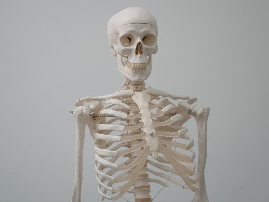
This lab introduces bone classification – long, short, flat, irregular, and sesamoid – alongside detailed bone structure examination․ Students will learn about compact and spongy bone, identifying key features like osteons and trabeculae․ The focus extends to skeletal system functions: support, protection, movement, mineral storage, and blood cell formation․
Bone Classification and Structure
This section details the five bone classifications: long, short, flat, irregular, and sesamoid, emphasizing structural differences․ Students will analyze compact and spongy bone tissues, identifying osteons, Haversian canals, and trabeculae․ Understanding bone composition – organic and inorganic – is crucial, alongside periosteum and endosteum functions․

Skeletal System Functions
Labs explore the skeletal system’s vital roles: support, protection, movement, mineral storage (calcium, phosphate), and blood cell formation (hematopoiesis)․ Students will investigate how bone structure enables these functions, relating bone markings to muscle attachments and joint articulation․ Analyzing case studies demonstrates functional implications of skeletal disorders․
Lab 6 & 7: Axial and Appendicular Skeleton
These labs focus on identifying and articulating the bones comprising the axial (skull, vertebral column, ribs, sternum) and appendicular (limbs, girdles) skeletons․ Students will differentiate bone features, analyze skeletal landmarks, and understand regional variations․ Practical exercises involve bone assembly and articulation, reinforcing anatomical relationships․
Skull Bones and Features
This lab meticulously examines cranial and facial bones, identifying sutures, foramina, and processes․ Students will differentiate between the frontal, parietal, temporal, occipital, sphenoid, and ethmoid bones, alongside the maxilla, mandible, and nasal bones․ Emphasis is placed on understanding the functional significance of specific skull features and their role in protecting vital structures․
Vertebral Column Regions and Characteristics
This lab focuses on identifying the cervical, thoracic, lumbar, sacral, and coccygeal regions of the vertebral column․ Students will analyze the typical and atypical vertebrae within each region, noting curvature, spinous process orientation, and intervertebral disc placement․ Understanding the structural adaptations supporting body weight and protecting the spinal cord is key․
Lab 8: Articulations – Joints and Movement
This lab explores the structural classification of joints, focusing on fibrous, cartilaginous, and synovial types․ Students will identify specific synovial joint features – articular cartilage, joint capsules, and ligaments – and correlate structure with range of motion․ Analyzing movements like flexion, extension, and rotation is crucial for understanding joint functionality․
Types of Synovial Joints
This section details the six classes of synovial joints: plane, hinge, pivot, condylar, saddle, and ball-and-socket․ Students will learn to differentiate these based on their shape and permitted movements․ Understanding how each joint type facilitates specific actions – like the elbow’s hinge motion or the shoulder’s wide range – is key․
Joint Movements and Range of Motion
This lab explores movements like flexion, extension, abduction, adduction, rotation, and circumduction, defining each with practical examples․ Range of motion, influenced by joint structure, ligaments, and muscle elasticity, will be assessed․ Students will analyze how these movements contribute to functional activities and understand limitations caused by joint injuries or diseases․
Lab 9: Muscles – Structure and Function
This lab investigates skeletal, smooth, and cardiac muscle tissues, detailing their microscopic anatomy and physiological roles․ Students will explore the sliding filament model, explaining muscle contraction mechanisms involving actin, myosin, and calcium ions․ Emphasis is placed on understanding how muscle structure dictates function, impacting movement, posture, and vital bodily processes․
Muscle Tissue Types
This section details skeletal, smooth, and cardiac muscle tissues, focusing on their unique structural and functional characteristics․ Skeletal muscle enables voluntary movement, while smooth muscle controls involuntary processes in internal organs․ Cardiac muscle, found exclusively in the heart, exhibits rhythmic contractions․ Students will analyze microscopic slides to differentiate these tissues․
Muscle Contraction Mechanism
This lab explores the sliding filament theory, detailing the roles of actin, myosin, and calcium ions in muscle contraction․ Students will investigate how nerve impulses trigger muscle fiber stimulation, leading to cross-bridge cycling and ultimately, muscle shortening․ Understanding this mechanism is crucial for comprehending movement and physiological processes․
Lab 10: Nervous System – Brain and Cranial Nerves
This lab focuses on identifying key brain regions and correlating them with their specific functions, enhancing neurological understanding․ Students will also learn to identify the twelve cranial nerves, detailing their origins, pathways, and associated sensory or motor functions․ This practical experience builds a foundation for clinical applications․
Brain Regions and Functions
This section details the major brain regions – cerebrum, cerebellum, and brainstem – and their roles in higher-order thinking, motor control, and vital life functions․ Students will explore the lobes of the cerebrum, associating each with specific sensory, motor, and cognitive processes․ Understanding these connections is crucial for neurological assessment․
Cranial Nerve Identification and Function
This lab focuses on identifying the twelve cranial nerves and correlating each with its specific sensory or motor function․ Students will learn to assess cranial nerve function through targeted clinical tests, understanding how deficits can indicate neurological damage․ Accurate identification is vital for diagnosing and treating various conditions․

Lab 11-16: Regional Muscle Anatomy
These labs detail the muscles of the shoulder, upper/lower arms, hip, thigh, leg, foot, head, and trunk․ Students will identify origins, insertions, actions, and innervations of key muscles․ Practical application involves palpation and movement analysis, solidifying understanding of regional muscular systems and their integrated functions within the human body․
Muscles of the Shoulder and Upper Arm
This section focuses on detailed anatomical study of muscles responsible for shoulder and arm movement․ Labs cover origins, insertions, actions, and innervations of deltoid, rotator cuff muscles, biceps brachii, triceps brachii, and brachialis; Students will learn functional relationships and apply knowledge through movement analysis and palpation exercises․
Muscles of the Forearm and Hand
This lab explores the intricate musculature controlling wrist, hand, and finger movements․ Students will dissect and identify flexors and extensors of the wrist and fingers, including muscles like flexor carpi ulnaris, extensor digitorum, and thenar/hypothenar eminences․ Emphasis is placed on understanding functional groups and their clinical relevance․
Muscles of the Hip and Thigh
This lab focuses on the powerful muscles responsible for locomotion and posture․ Students will dissect and identify major hip flexors, extensors, abductors, and adductors, alongside thigh muscles like the quadriceps and hamstrings․ Understanding origins, insertions, and actions is crucial, alongside clinical correlations like strains and injuries․
Muscles of the Leg and Foot
This lab explores the intricate musculature enabling walking, running, and maintaining balance․ Students will dissect and identify anterior, posterior, and lateral leg compartments, including muscles like the tibialis anterior, gastrocnemius, and fibularis longus․ Emphasis is placed on understanding foot movements, arches, and common injuries like sprains and plantar fasciitis․
Muscles of the Head and Neck
This lab focuses on the complex muscle groups controlling facial expressions, chewing, and head movements․ Students will identify muscles like the masseter, temporalis, sternocleidomastoid, and various facial muscles․ Dissection and palpation exercises will demonstrate origins, insertions, and actions, alongside clinical correlations like temporomandibular joint (TMJ) disorders․
Muscles of the Trunk
This lab explores muscles vital for posture, movement, and respiration, including abdominal, back, and intercostal muscles․ Students will dissect and identify muscles like the rectus abdominis, external and internal obliques, erector spinae, and diaphragm․ Emphasis will be placed on understanding their roles in core stability, spinal support, and breathing mechanics․
Lab 17: Special Senses – Taste and Smell
This lab investigates chemoreception, focusing on taste receptor distribution across the tongue and the mechanism of olfactory receptor activation within the nasal cavity․ Students will explore how these senses contribute to flavor perception and environmental awareness, examining the neural pathways involved in transmitting sensory information to the brain․
Taste Receptor Distribution and Function

This section details the anatomy of taste buds, primarily located on papillae, and their role in detecting five primary tastes: sweet, sour, salty, bitter, and umami․ Students will analyze regional variations in receptor sensitivity, debunking the traditional “tongue map,” and understand how taste interacts with olfactory senses to create flavor perception․
Olfactory Receptor Location and Mechanism
This lab explores the olfactory epithelium within the nasal cavity, housing millions of olfactory receptor neurons․ Students will investigate how odorant molecules bind to these receptors, triggering a signal transduction cascade leading to brain interpretation․ The process of adaptation and the connection between smell and memory will also be examined․
Lab 18: Eye and Ear – Vision and Hearing
This lab dissects the anatomy of the eye, tracing the visual pathway from the retina to the brain, and explores how light is focused for clear vision․ Simultaneously, students will examine the ear’s structures, detailing the auditory pathway and mechanisms responsible for hearing and maintaining balance, crucial sensory functions․
Anatomy of the Eye and Visual Pathway
This section meticulously details the eye’s components – cornea, lens, retina, and optic nerve – and their roles in vision․ Students will trace the visual pathway, understanding how signals travel from photoreceptors to the brain for interpretation, enabling sight․ Stunning photographs and illustrations enhance comprehension of these complex structures․
Anatomy of the Ear and Auditory Pathway
This lab explores the ear’s intricate anatomy – outer, middle, and inner ear – and their function in hearing and balance․ Students will map the auditory pathway, tracing sound wave transmission from the tympanic membrane through ossicles to the cochlea, and ultimately to the auditory cortex for processing․